Small Bowel Cancer Samples
Bay Biosciences provides high quality, clinical grade, fresh frozen tumor tissue bio-specimens, cryogenically preserved sera (serum), plasma and peripheral blood mononuclear cells (PBMC) biofluid samples from patients diagnosed with Small Bowel Cancer.
The sera (serum), plasma and PBMC biofluid specimens are processed from patient’s peripheral whole-blood using customized collection and processing protocols. The small bowel cancer fresh frozen tumor tissue samples and biofluids are collected from unique patients diagnosed with small bowel cancer and are provided to a valued pharmaceutical customer for research, diagnostics, discovery and drug development.
Detailed clinical data, small bowel cancer patients history, symptoms, complete blood count (CBC), serology, chemotherapy information, tumor biopsy tissue blocks, elevated biomarker levels, genetic and metabolic information, histopathological findings, annotations associated with small bowel cancer specimens is provided to a valued customer for research, development and drug discovery.
The small bowel cancer sera (serum), plasma and peripheral blood mononuclear cells (PBMC) biofluid are processed from patients peripheral whole-blood using customized collection and processing protocols.

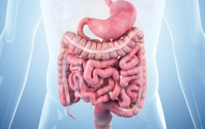
The Small Bowel
The small bowel is part of the digestive system. Its function is to break down food and nutrients to be absorbed into the body. The small bowel is also called the small intestine. It links the stomach to the large intestine, which is known as the colon. The small bowel is divided into the following three parts:
- The Duodenum: The part closest to the stomach
- The Jejunum: The middle portion
- The Ileum: The bottom section, which connects to the large intestine, or colon.
The small bowel is approximately 15 feet long, folds many times to fit inside the abdomen, and makes up about seventy percent of the entire digestive system and ninety percent of the absorptive surface area of the gastrointestinal tract.
Small Bowel Cancer
Small bowel cancer starts when healthy cells in the lining of the small bowel change and grow out of control, forming a mass called a tumor. A tumor can be cancerous or benign. A cancerous tumor is malignant, meaning it can grow and spread to other parts of the body. A benign tumor means that the tumor can grow but will not spread. These changes can take a long time to develop. Both genetic and environmental factors can cause such changes, although the specific causes of small bowel cancer are generally not well understood. Although the small bowel represents 90% of the surface area and 75% of the length of the alimentary tract and is located between two organs with high cancer incidence (i.e., stomach and colon), malignant neoplasm of the small bowel fall in the category of rare neoplasms. They account for only 2% of all GI malignancies.
Types of Small Bowel Cancer
There are Five Main Types of Small Bowel Cancer’s:
- Adenocarcinoma: Adenocarcinoma is the most common type of small bowel cancer, usually occurring in the duodenum or jejunum. Adenocarcinoma begins in the gland cells of the small bowel.
- Sarcoma: Small bowel sarcoma is generally a leiomyosarcoma, which is a tumor that arises in the muscle tissue that makes up part of the intestine. This type of tumor most often occurs in the ileum.
- Gastrointestinal stromal tumor (GIST): GIST is an uncommon tumor that is believed to start in cells found in the walls of the gastrointestinal (GI) tract, called interstitial cells of Cajal (ICC). GIST belongs to a group of cancers called soft tissue sarcomas.
- Neuroendocrine tumor: These can also be called a carcinoid tumor. These are tumors that start in the hormone-producing cells of various organs and generally occur in the ileum.
- Lymphoma: Lymphoma is a cancer of the lymph system, which is part of the body’s immune system. Lymphoma that occurs in the small bowel usually occurs in the jejunum or ileum and is most commonly non-Hodgkin lymphoma.
Causes of Small Bowel Cancer
Exact causes of most small bowel cancers are unknown Generally small bowel cancer begins when healthy cells in the small bowel develop changes (mutations) in their DNA. A cell’s DNA contains a set of instructions that tell a cell what to do. Healthy cells grow and divide in an orderly way to keep your body functioning normally. But when a cell’s DNA is damaged and becomes cancerous, cells continue to divide, even when new cells aren’t needed. As these cells accumulate, they form a tumor. Over time, the cancer cells can grow to invade and destroy normal tissue nearby. And cancerous cells can spread (metastasize) to other parts of the body.
Signs and Symptoms of Small Bowel Cancer
Patients with small bowel cancer may experience the following symptoms or signs. Sometimes, patients with small bowel cancer do not have any of these changes. Or, the cause of a symptom may be a different medical condition that is not cancer.
- Abdominal pain
- A lump in the abdomen
- Blood in the stool (feces)
- Dark colored (black) stools
- Episodes of abdominal pain that may be accompanied by severe nausea or vomiting
- Fatigue
- Nausea
- Pain or cramps in the abdomen
- Skin flushing
- Unexplained weight loss
- Vomiting
- Watery Diarrhea
- Yellowing of the skin and the whites of the eyes (jaundice)
Risk Factors of Small Bowel Cancer
The following factors may raise a person’s risk of small bowel adenocarcinoma:
- Crohn’s Disease: Crohn’s disease is a chronic inflammation of the gastrointestinal tract. Patients with Crohn’s disease have a higher risk of both Colorectal cancer and small bowel adenocarcinomas.
- Celiac disease: Celiac disease is a digestive disease that interferes with the absorption of nutrients from food in the small bowel. The body’s immune system responds to a protein mixture called gluten, which is found in wheat, rye, barley, oats, and other grain foods and can damage the lining of the small bowel.
- Familial adenomatous polyposis (FAP): FAP is an inherited condition characterized by hundreds or thousands of colon polyps, which are small growths. The polyps are usually benign (noncancerous), but there is nearly a 100% chance that the polyps will develop into cancer if left untreated. Individuals with FAP are also at risk for other types of cancer, including stomach cancer, duodenal cancer, thyroid cancer, pancreatic cancer, and hepatoblastoma, which is liver cancer seen mainly in early childhood.
Diagnosis of Small Bowel Cancer
In addition to a physical examination, following diagnostics tests may be used to diagnose small bowel cancer:
- Blood Tests: A test of the number of red blood cells in the blood can indicate whether the cancer is causing any bleeding. Tests for your liver and kidney function may also be performed. The results will determine if either of those organs may be affected by the cancer and find out how healthy those organs are before having treatment for small bowel cancer.
- X-ray: An x-ray is way to create a picture of the structures inside of the body using a small amount of radiation. It can help the doctor find a tumor. For small bowel cancer, x-rays may be taken of the entire gastrointestinal system, including the esophagus, stomach, small bowel, large intestine, and rectum. Sometimes, the person will drink a substance called barium, which outlines the esophagus, stomach, and small bowel on the x-ray and helps the doctor see tumors or other abnormal areas. This is called an upper gastrointestinal series with small bowel follow-through (UGI SBFT). To get a better picture of the lower gastrointestinal tract, a barium enema may be performed. In this procedure, barium is placed into the rectum and coats the rectum and large intestine. Abdominal x-rays may also show the location of a tumor.
- Biopsy: A biopsy is the removal of a small amount of tissue for examination under a microscope. Other tests can suggest that cancer is present, but only a biopsy can make a definite diagnosis. A pathologist then analyzes the sample(s). A pathologist is a doctor who specializes in interpreting laboratory tests and evaluating cells, tissues, and organs to diagnose disease.
- Endoscopy: An endoscopy allows the doctor to see the inside the gastrointestinal system. The person may be sedated while the doctor inserts a thin, lighted, flexible tube called an endoscope through the mouth, down the esophagus, and into the stomach and small bowel. Sedation is giving medication to become more relaxed, calm, or sleepy. If abnormal areas are found, the doctor can remove a sample of tissue and check it for evidence of cancer. An endoscopy allows the doctors to see some, but not all, of the small bowel. Because of this, the doctor usually recommends a videocapsule endoscopy (VCE). In this method, the patient swallows a small (pill-sized) capsule that contains a tiny camera and light. Pictures are collected from the capsule as it travels through the patient’s gastrointestinal system. The capsule exits the body during the patient’s next bowel movement.
- Colonoscopy: A colonoscopy is similar to the traditional endoscopy described above, except that the endoscope enters the body through the anus and rectum into the colon and lower part of the small bowel.
- Computed tomography (CT or CAT) scan: A CT scan take pictures of the inside of the body using x-rays taken from different angles. A computer combines these images into a detailed, 3-dimensional or 3-D image that shows any abnormalities or tumors. A CT scan can be used to measure the tumor’s size. Sometimes, a special dye called a contrast medium is given before the scan to provide better detail on the image. This dye can be injected into a patient’s vein or given as a pill or liquid to swallow. A CT scan can check for the spread of cancer to the lungs, liver, and other organs.
- Positron emission tomography (PET) or PET-CT scan: A PET scan is usually combined with a CT scan called a PET-CT scan. A PET scan is a way to create pictures of organs and tissues inside the body. A small amount of a radioactive sugar substance is injected into the patient’s body. This sugar substance is taken up by cells that use the most energy. Because cancer tends to use energy actively, it absorbs more of the radioactive substance. A scanner detects this substance to produce images of the inside of the body.
- Laparotomy: In this procedure, a surgical incision is made in the abdomen to check for cancer and tumor’s. Sometimes, tissue samples are taken and, often, surgery is performed at the same time to remove the tumor.

Bay Biosciences is a global leader in providing researchers with high quality, clinical grade, fully characterized human tissue samples, bio-specimens and human bio-fluid collections from cancer (tumor) tissue, cancer serum, cancer plasma cancer PBMC and human tissue samples from most other therapeutic areas and diseases.
Bay Biosciences maintains and manages it’s own bio-repository, human tissue bank (biobank) consisting of thousands of diseased samples (specimens) and from normal healthy donors available in all formats and types. Our biobank procures and stores fully consented, deidentified and institutional review boards (IRB) approved human tissue samples and matched controls.
All our human human tissue collections, human specimens and human bio-fluids are provided with detailed samples associated patient’s clinical data. This critical patient’s clinical data includes information relating to their past and current disease, treatment history, lifestyle choices, biomarkers and genetic information. Patient’s data is extremely valuable for researchers and is used to help identify new effective treatments (drug discovery & development) in oncology, other therapeutic areas and diseases. This clinical information is critical to demonstrate their impact, monitor the safety of medicines, testing & diagnostics, and generate new knowledge about the causes of disease and illness.
Bay Biosciences banks wide variety of human tissue samples and biological samples including cryogenically preserved -80°C, fresh, fresh frozen tissue samples, tumor tissue samples, FFPE’s, tissue slides, with matching human bio-fluids, whole blood and blood derived products such as serum, plasma and PBMC’s.
Bay Biosciences is a global leader in collecting and providing human tissue samples according to the researchers specified requirements and customized, tailor made collection protocols. Please contact us anytime to discuss your special research projects and customized human tissue sample requirements.
Bay Biosciences provides human tissue samples (human specimens) from diseased and normal healthy donors; including peripheral whole-blood, amniotic fluid, bronchoalveolar lavage fluid (BAL), sputum, pleural effusion, cerebrospinal fluid (CSF), serum (sera), plasma, peripheral blood mononuclear cells (PBMC’s), saliva, Buffy coat, urine, stool samples, aqueous humor, vitreous humor, kidney stones, renal calculi, nephrolithiasis, urolithiasis and other bodily fluids from most diseases including cancer. We can also procure most human bio-specimens and can do special collections and requests of human samples that are difficult to find. All our human tissue samples are procured through IRB approved clinical protocols and procedures.
In addition to the standard processing protocols Bay Biosciences can also provide human plasma, serum, PBMC bio-fluid samples using custom processing protocols, you can buy donor specific sample collections in higher volumes and specified sample aliquoting from us. Bay Biosciences also provides human samples from normal healthy donors, volunteers, for controls and clinical research, contact us Now.
日本のお客様は、ベイバイオサイエンスジャパンBay Biosciences Japanまたはhttp://baybiosciences-jp.com/contact/までご連絡ください。


