Cerebrovascular Disease Samples
Bay Biosciences provides high quality, clinical grade, bio-specimens and matched cryogenically preserved sera (serum), plasma and peripheral blood mononuclear cells (PBMC) biofluid samples from patients diagnosed with cerebrovascular disease.
The sera (serum), plasma and PBMC biofluid specimens are processed from cerebrovascular disease patient’s peripheral whole-blood using customized collection and processing protocols. The cerebrovascular disease biofluid samples are collected from unique patients diagnosed with cerebrovascular disease and are provided to a valued pharmaceutical customer for research, diagnostics, discovery and drug development.
Detailed clinical data, patients history, symptoms, complete blood count (CBC), serology, MRI, CT scan, histopathology information, elevated biomarker levels, genetic and metabolic information associated with cerebrovascular disease specimens is provided to a valued customer for research, development and drug discovery.
The cerebrovascular disease sera (serum), plasma and peripheral blood mononuclear cells (PBMC) biofluids are processed from patients peripheral whole-blood using customized collection and processing protocols.

Cerebrovascular Disease Overview
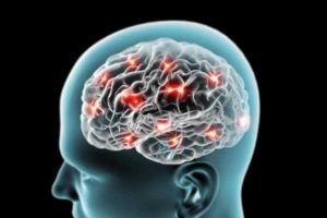
Cerebrovascular disease refers to a group of conditions, diseases, and disorders that affect the blood vessels and blood supply to the brain. Cerebrovascular disease includes stroke, carotid stenosis, vertebral stenosis and intracranial stenosis, aneurysms and vascular malformations. Restrictions in blood flow may occur from vessel narrowing (stenosis), clot formation (thrombosis), blockage (embolism) or blood vessel rupture (hemorrhage). Lack of sufficient blood flow (ischemia) affects brain tissue and may cause a stroke.
The word cerebrovascular is made up of two parts “cerebro” which refers to the large part of the brain, and “vascular” which means arteries and veins. Together, the word cerebrovascular refers to blood flow in the brain. The term cerebrovascular disease includes all disorders in which an area of the brain is temporarily or permanently affected by ischemia or bleeding and one or more of the cerebral blood vessels are involved in the pathological process.
Cerebrovascular disease is the fifth most common cause of death in the United States. According to the Centers for Disease Control and Prevention (CDC) cerebrovascular disease causes over 150,000 deaths per year or 45.7deaths per 100,000 population.
Blood Flow to the Brain
The brain relies on only two sets of major arteries for its blood supply, it is very important that these arteries are healthy. Often, the underlying cause of an ischemic stroke is carotid arteries blocked with a fatty buildup, called plaque. During a hemorrhagic stroke, an artery in or on the surface of the brain has ruptured or leaks, causing bleeding and damage in or around the brain. Without oxygen and important nutrients, the affected brain cells are either damaged or die within a few minutes. Once brain cells die, they cannot regenerate, and devastating damage may occur, sometimes resulting in physical, cognitive and mental disabilities.
Types of Cerebrovascular Disease
Cerebrovascular diseases types include stroke, transient ischemic attack (TIA), aneurysm, vascular malformation, and subarachnoid hemorrhage. Aneurysms and hemorrhages might cause severe health problems. Blood clots can form in the brain or travel there from other parts of the body, causing a blockage.
Following are the different types of cerebrovascular diseases:
Ischemic stroke: Ischemic stroke occurs when a blood clot or atherosclerotic plaque blocks a blood vessel that supplies blood to the brain. A clot, or thrombus, may form in an artery that is already narrow. A stroke happens when the lack of blood supply results in the death of brain cells.
Embolism: An embolism occurs when a clot breaks off from elsewhere in the body and travels to the brain to block a smaller artery. Embolism or an embolic stroke is the most common type of ischemic stroke. Patients who have arrhythmias, which are conditions that cause an irregular heart rhythm, are more prone to developing an embolism. A tear in the lining of the carotid artery, which is in the neck, can lead to ischemic stroke. The tear lets blood flow between the layers of the carotid artery, narrowing it, and reducing blood supply to the brain.
Hemorrhagic stroke: Hemorrhagic stroke (bleeds) occurs when a blood vessel in part of the brain weakens and bursts open, causing blood to leak into the brain. The leaking blood puts pressure on the brain tissue, leading to edema, which damages brain tissue. The hemorrhage can also cause nearby parts of the brain to lose their supply of oxygen rich blood.
Cerebral aneurysm or subarachnoid hemorrhage: An aneurysm is a bulge in the arterial wall that can rupture and bleed. Cerebral aneurysm can result from structural problems in the blood vessels of the brain. A subarachnoid hemorrhage occurs when a blood vessel ruptures and bleeds between two membranes surrounding the brain. This leaking of blood can damage brain cells.
Signs and Symptoms of Cerebrovascular Disease
Signs and symptoms of cerebrovascular disease depend on the location of the blockage and its impact on brain tissue. Different events may have different effects. Following are the common signs and symptoms of cerebrovascular disease:
- Confusion and disorientation
- Unconsciousness
- Communications problems
- Headaches, (severe and sudden)
- Hemiplegia (paralysis of one side of the body)
- Hemiparesis (weakness on one side of the body)
- Losing vision (on one side)
- Loss of balance
- Slurred speech
Causes of Cerebrovascular Disease
There are several causes for developing cerebrovascular disease develops. If damage occurs to a blood vessel in the brain, it will not be able to deliver enough or any blood to the area of the brain that it serves. The lack of blood interferes with the delivery of adequate oxygen, and, without oxygen, brain cells will start to die. Brain damage is irreversible. Emergency help is vital to reduce a patients risk of long term brain damage and increase their chances of survival. Atherosclerosis is a primary cause of cerebrovascular disease. This occurs when high cholesterol levels, together with inflammation in the arteries of the brain, cause cholesterol to build up as a thick, waxy plaque that can narrow or block blood flow in the arteries. This plaque can limit or completely obstruct blood flow to the brain, causing a cerebrovascular attack, such as a stroke or transient ischemic attack (TIA).
Diagnosis of Cerebrovascular Disease
The majority of cerebrovascular diseases can be identified through diagnostic imaging tests. These tests allow neurosurgeons to view the arteries and vessels in and around the brain and the brain tissue itself. Following are some of the diagnostic tests listed below used to diagnose cerebrovascular diseases:
Cerebral angiography (also called vertebral angiogram, carotid angiogram): Arteries are not normally seen in an X-ray, so contrast dye is utilized. The patient is given a local anesthetic, the artery is punctured, usually in the leg, and a needle is inserted into the artery. A catheter (a long, narrow, flexible tube) is inserted through the needle and into the artery. It is then threaded through the main vessels of the abdomen and chest until it is properly placed in the arteries of the neck. This procedure is monitored by a fluoroscope (a special X-ray that projects the images on a TV monitor). The contrast dye is then injected into the neck area through the catheter and X-ray pictures are taken.
Carotid duplex (also called carotid ultrasound): In this procedure, ultrasound is used to help detect plaque, blood clots or other problems with blood flow in the carotid arteries. A water-soluble gel is placed on the skin where the transducer (a handheld device that directs the high-frequency sound waves to the arteries being tested) is to be placed. The gel helps transmit the sound to the skin surface. The ultrasound is turned on and images of the carotid arteries and pulse wave forms are obtained. There are no known risks and this test is noninvasive and painless.
Computed tomography (CT or CAT scan): A diagnostic image created after a computer reads x-rays. In some cases, a medication will be injected through a vein to help highlight brain structures. Bone, blood and brain tissue have very different densities and can easily be distinguished on a CT scan. A CT scan is a useful diagnostic test for hemorrhagic strokes because blood can easily be seen. However, damage from an ischemic stroke may not be revealed on a CT scan for several hours or days and the individual arteries in the brain cannot be seen. CTA (CT angiography) allows clinicians to see blood vessels of the head and neck and is increasingly being used instead of an invasive angiogram.
Doppler ultrasound: A water-soluble gel is placed on the transducer (a handheld device that directs the high-frequency sound waves to the artery or vein being tested) and the skin over the veins of the extremity being tested. There is a “swishing” sound on the Doppler if the venous system is normal. Both the superficial and deep venous systems are evaluated. There are no known risks and this test is noninvasive and painless.
Electroencephalogram (EEG): A diagnostic test using small metal discs (electrodes) placed on a person’s scalp to pick up electrical impulses. These electrical signals are printed out as brain waves.
Lumbar puncture (spinal tap): An invasive diagnostic test that uses a needle to remove a sample of cerebrospinal fluid from the space surrounding the spinal cord. This test can be helpful in detecting bleeding caused by a cerebral hemorrhage.
Magnetic Resonance Imaging (MRI): A diagnostic test that produces three-dimensional images of body structures using magnetic fields and computer technology. It can clearly show various types of nerve tissue and clear pictures of the brain stem and posterior brain. An MRI of the brain can help determine whether there are signs of prior mini-strokes also known as transient ischemic attack (TIA). This test is noninvasive, although some patients may experience claustrophobia in the imager.
Magnetic Resonance Angiogram (MRA): This is a noninvasive test which is conducted in a Magnetic Resonance Imager (MRI). The magnetic images are assembled by a computer to provide an image of the arteries in the head and neck. The MRA shows the actual blood vessels in the neck and brain and can help detect blockage and aneurysms in the body.

Bay Biosciences is a global leader in providing researchers with high quality, clinical grade, fully characterized human tissue samples, bio-specimens and human bio-fluid collections from cancer (tumor) tissue, cancer serum, cancer plasma cancer PBMC and human tissue samples from most other therapeutic areas and diseases.
Bay Biosciences maintains and manages it’s own bio-repository, human tissue bank (biobank) consisting of thousands of diseased samples (specimens) and from normal healthy donors available in all formats and types. Our biobank procures and stores fully consented, deidentified and institutional review boards (IRB) approved human tissue samples and matched controls.
All our human human tissue collections, human specimens and human bio-fluids are provided with detailed samples associated patient’s clinical data. This critical patient’s clinical data includes information relating to their past and current disease, treatment history, lifestyle choices, biomarkers and genetic information. Patient’s data is extremely valuable for researchers and is used to help identify new effective treatments (drug discovery & development) in oncology, other therapeutic areas and diseases. This clinical information is critical to demonstrate their impact, monitor the safety of medicines, testing & diagnostics, and generate new knowledge about the causes of disease and illness.
Bay Biosciences banks wide variety of human tissue samples and biological samples including cryogenically preserved -80°C, fresh, fresh frozen tissue samples, tumor tissue samples, FFPE’s, tissue slides, with matching human bio-fluids, whole blood and blood derived products such as serum, plasma and PBMC’s.
Bay Biosciences is a global leader in collecting and providing human tissue samples according to the researchers specified requirements and customized, tailor made collection protocols. Please contact us anytime to discuss your special research projects and customized human tissue sample requirements.
Bay Biosciences provides human tissue samples (human specimens) from diseased and normal healthy donors; including peripheral whole-blood, amniotic fluid, bronchoalveolar lavage fluid (BAL), sputum, pleural effusion, cerebrospinal fluid (CSF), serum (sera), plasma, peripheral blood mononuclear cells (PBMC’s), saliva, Buffy coat, urine, stool samples, aqueous humor, vitreous humor, kidney stones, renal calculi, nephrolithiasis, urolithiasis and other bodily fluids from most diseases including cancer. We can also procure most human bio-specimens and can do special collections and requests of human samples that are difficult to find. All our human tissue samples are procured through IRB approved clinical protocols and procedures.
In addition to the standard processing protocols Bay Biosciences can also provide human plasma, serum, PBMC bio-fluid samples using custom processing protocols, you can buy donor specific sample collections in higher volumes and specified sample aliquoting from us. Bay Biosciences also provides human samples from normal healthy donors, volunteers, for controls and clinical research, contact us Now.
日本のお客様は、ベイバイオサイエンスジャパンBay Biosciences Japanまたはhttp://baybiosciences-jp.com/contact/までご連絡ください。


